
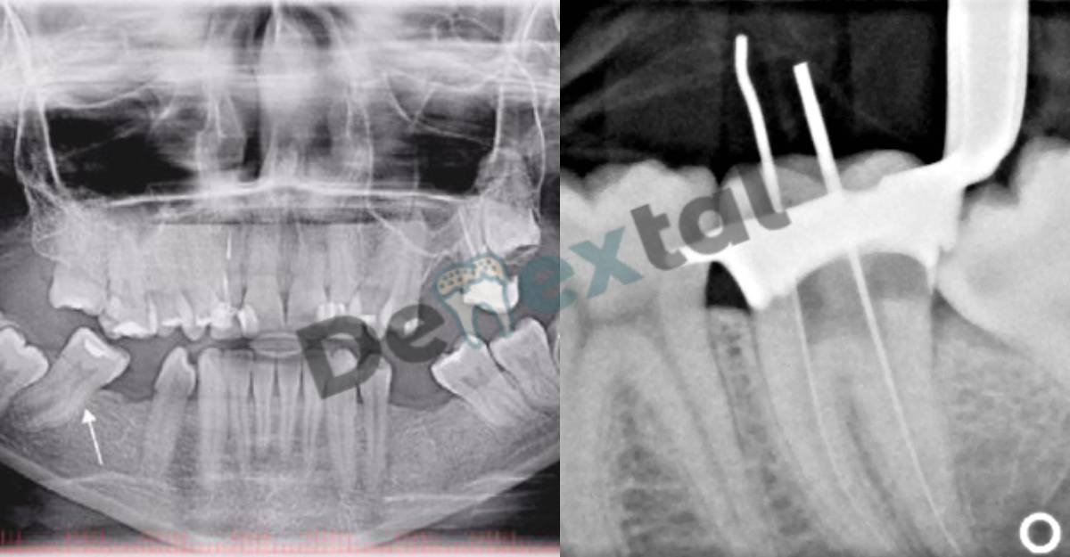
A Root Canal X-Rays, also known as an endodontic radiograph, is a special type of dental x-ray that is used to take detailed images of the inside of a tooth, specifically the root canals. This procedure is typically done when a dentist or endodontist suspects that a patient may have an infection or inflammation in their tooth’s pulp, which is the soft tissue inside the tooth that contains nerves and blood vessels. This type of infection, known as pulpal necrosis, can cause severe pain and lead to the loss of the tooth if left untreated.
How is a Root Canal X-Ray performed?
During a Root Canal X-Ray, the patient will be seated in a dental chair and will have a small device, called a bite-wing, placed in their mouth to hold their jaw open. The dentist or endodontist will then use a special x-ray machine to take multiple images of the affected tooth from different angles. The patient will need to stay still during the procedure to ensure that the images are clear. The x-ray will show the shape, number and position of the root canals, as well as any signs of infection, such as bone loss or cysts.
Benefits of Root Canal X-Rays
Root Canal X-Rays can help dentists and endodontists to diagnose and treat a variety of dental problems, including infections, inflammation, and abscesses. They can also be used to determine the shape and size of the root canals, which is important for planning and performing root canal therapy. Additionally, Root Canal X-Rays can help to identify any other issues in the surrounding teeth or jaw that may be contributing to the patient’s dental problems. This type of x-ray also allows the dentist to check for any additional canals, which can be missed during a visual examination.
Risks of Root Canal X-Rays
As with any x-ray procedure, there is a small amount of radiation exposure associated with Root Canal X-Rays. However, the radiation levels are considered to be very low and the procedure is considered to be safe for most patients. The risk of developing cancer from a single Root Canal X-Ray is considered to be extremely low. It is also important to note that modern digital x-ray machines emit less radiation compared to traditional film-based machines.

Frequently Asked Questions
How often can I get a Root Canal X-Ray?
As the procedure is considered safe, the frequency of Root Canal X-ray is determined by the dentist or endodontist. It depends on the diagnosis and treatment plan.
Is a Root Canal X-Ray painful?
No, the procedure is not painful. There may be some discomfort when the device is placed in the mouth to hold the jaw open.
How Are The results of a Root Canal X-Ray interpreted?
The dentist or endodontist will look at the images taken during the Root Canal X-Ray to diagnose any dental problems, plan treatment, and determine the shape and size of the root canals. They will also look for any signs of infection or inflammation, such as bone loss or cysts. The results of the x-ray will be used to guide the dentist or endodontist in the development of an appropriate treatment plan, which may include root canal therapy or extraction.
Conclusion
Root Canal X-Rays are a valuable tool for dentists and endodontists in the diagnosis and treatment of dental problems. The procedure is safe, quick, and non-invasive, and it provides detailed images of the inside of a tooth that can help to identify issues such as infections and inflammation. If you suspect that you may have a problem with one of your teeth, be sure to speak with your dentist or endodontist about the possibility of a Root Canal X-Ray.